
greeshma-suresh

Dr. Greeshma Suresh

Pediatric Surgeon

M.Ch (Pediatric Surgery), M.S (General Surgery), MBBS

6+ Years of Experience

1500+ surgical cases managed

6+ Years of Experience

1500+ surgical cases managed

Vatsalya Children Hospital
LinQ Card

About Dr. Greeshma Suresh

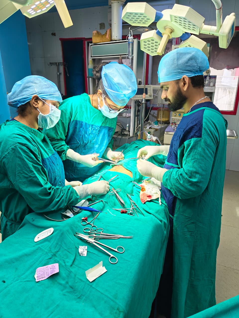
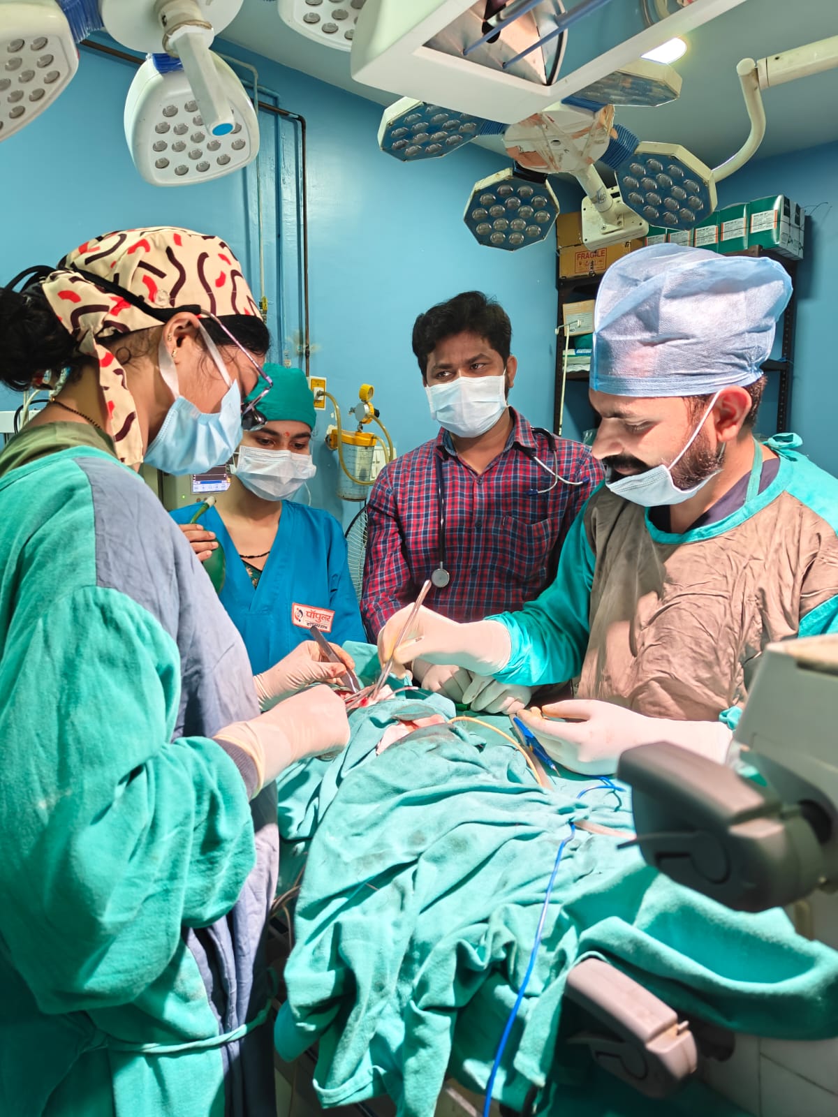
Dr. Greeshma Suresh is a highly skilled and accomplished Pediatric Surgeon with over 6 years of extensive clinical and surgical experience, specializing in advanced neonatal, infant, and pediatric surgical care. With an impressive track record of managing 1500+ complex surgical cases, Dr. Greeshma Suresh is renowned for precision-driven techniques, compassionate patient care, and excellent clinical outcomes. Her expertise spans minimally invasive pediatric surgery, congenital anomaly corrections, pediatric urology, and emergency surgical management. Trained at prestigious institutions, including the Institute of Medical Sciences, Banaras Hindu University, she combines academic excellence with hands-on surgical mastery. Dr. Greeshma Suresh is also an active contributor to medical research with multiple national and international publications, reflecting her commitment to innovation and evidence-based practice. Dedicated to delivering holistic, child-centric care, she ensures personalized treatment plans that prioritize safety, faster recovery, and long-term well-being, making her a trusted name in pediatric surgical excellence.


https://d2vvc091k8tmib.cloudfront.net/s3fs-public/2026-04/Dr.%20Greeshma%20overview.jpeg

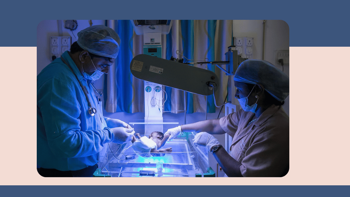
Dr. Greeshma Suresh specializes in Pediatric Surgery with a strong focus on delivering advanced, safe, and child-centric surgical care for neonates, infants, and children. Her core expertise includes neonatal surgeries for congenital anomalies such as anorectal malformations, tracheoesophageal fistula, intestinal atresia, and congenital diaphragmatic hernia, along with pediatric gastrointestinal and urological procedures like appendicectomy, cholecystectomy, herniotomy, undescended testes correction, hypospadias repair, and hydronephrosis management. She is highly skilled in minimally invasive (laparoscopic) pediatric surgery, ensuring reduced pain, faster recovery, and minimal scarring. Dr. Greeshma Suresh also excels in managing pediatric surgical emergencies, including acute abdomen and trauma cases, with precision and efficiency. With a patient-first approach and evidence-based practice, she is committed to providing comprehensive pediatric surgical treatment, making her a trusted expert for complex and routine surgical conditions in children.


Dr. Greeshma Suresh 's Areas of expertise

  • Neonatal Surgery: Expert in managing complex congenital anomalies in newborns with precision and compassionate care.
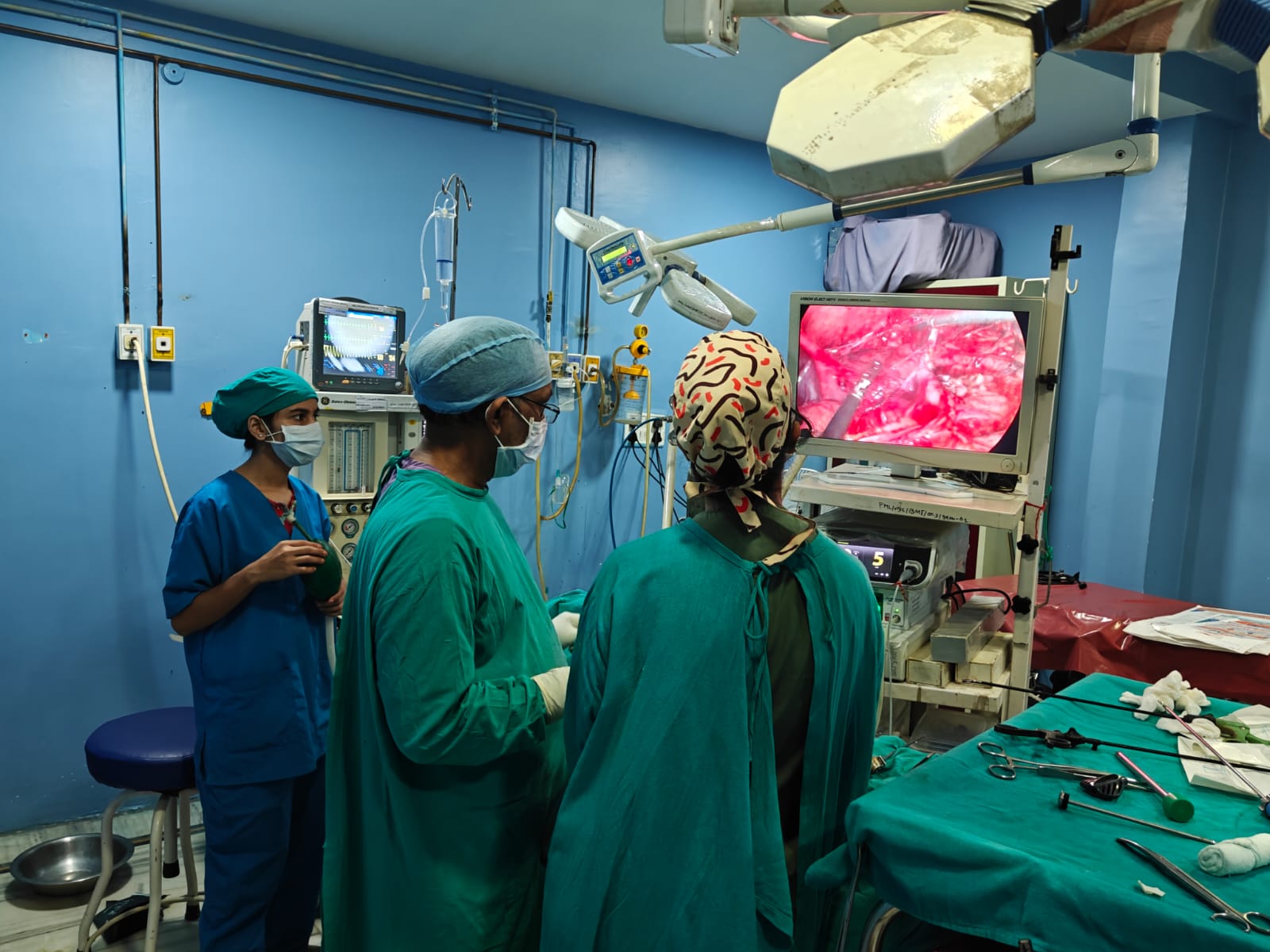
  • Pediatric Laparoscopic Surgery: Skilled in minimally invasive procedures ensuring faster recovery and minimal scarring.
  • Anorectal Malformations Treatment: Specialized in corrective surgeries for congenital anorectal disorders in infants and children.
  • Tracheoesophageal Fistula (TEF) Repair: Proficient in managing and surgically correcting TEF with high success outcomes.
  • Pediatric Urology Procedures: Experienced in treating conditions like undescended testes, hypospadias, and hydronephrosis.
  • Appendicectomy in Children: Expertise in safe and effective surgical removal of the appendix in pediatric patients.
  • Herniotomy: Skilled in performing hernia repairs in infants and children with excellent postoperative outcomes.
  • Management of Pediatric Trauma: Efficient in handling emergency surgical cases including abdominal and thoracic injuries.
  • Congenital Diaphragmatic Hernia Repair: Specialized in life-saving surgical correction of diaphragmatic defects in newborns.
  • Hirschsprung Disease Management: Experienced in diagnosing and surgically treating intestinal motility disorders in children.
Awards:
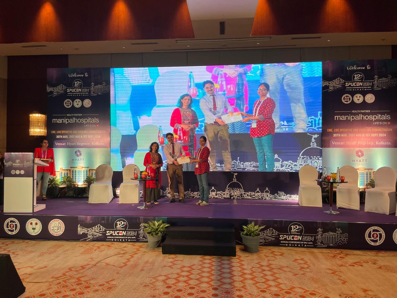
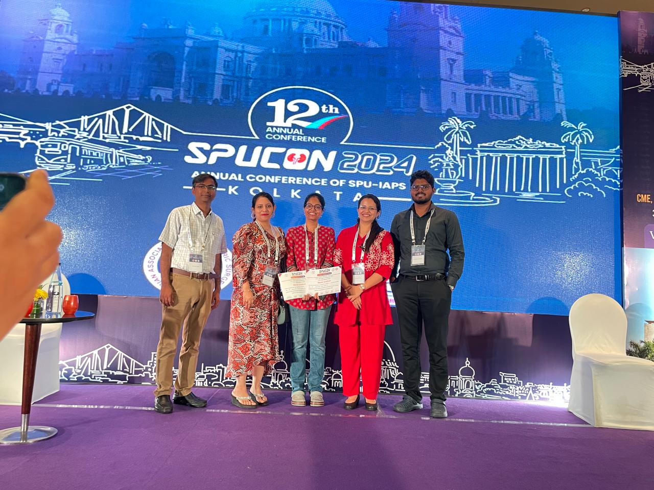
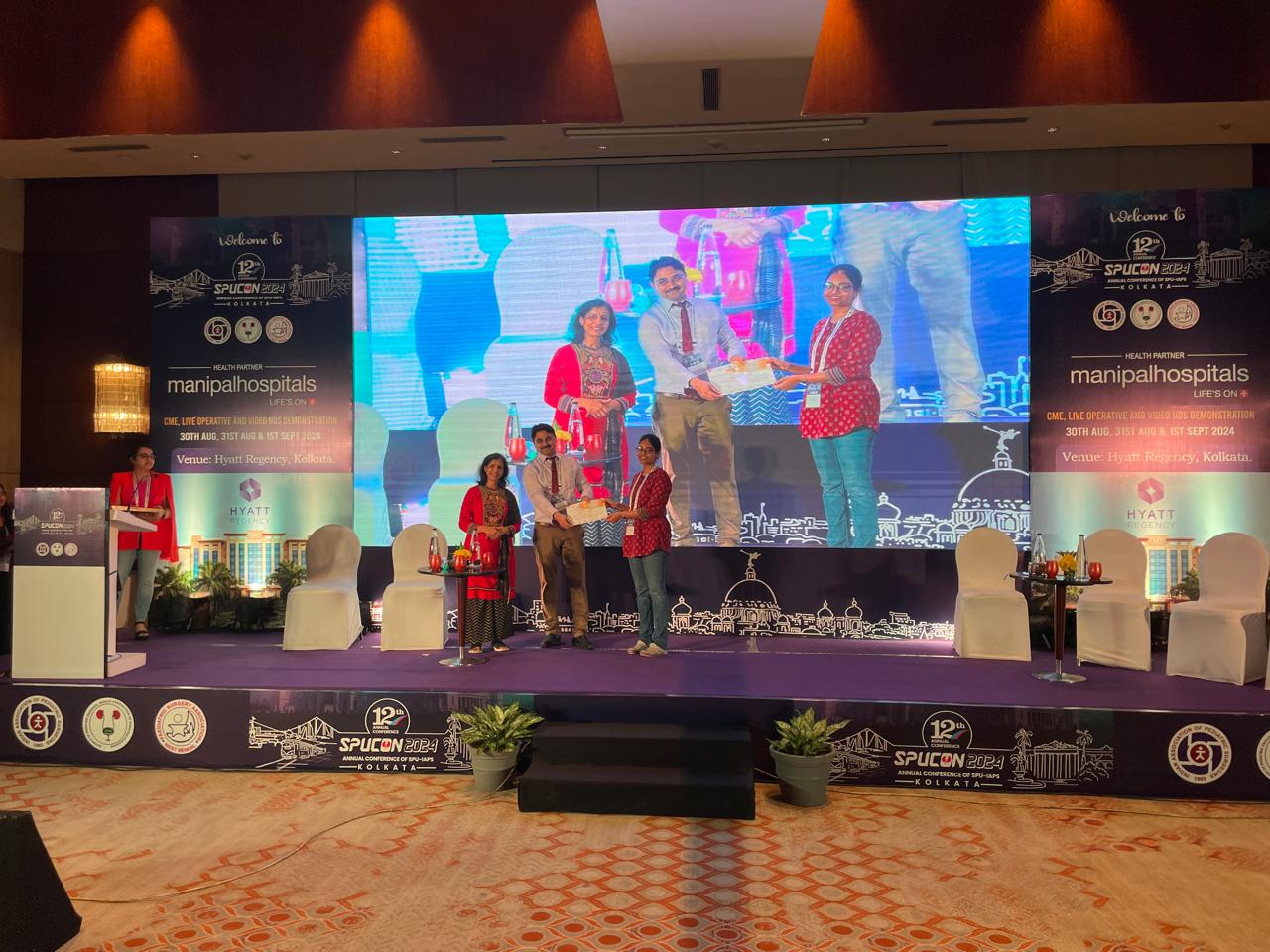
  • 1st Prize – Best Paper Award at SPUCON 2024
  • 1st Prize – Best Poster Award at SPUCON 2024
Publications:
  • Suresh G et al. Aldosterone and Renin as Prognostic Biomarkers in Posterior Urethral Valve – Iranian Journal of Pediatric Surgery (2024)
  • Suresh G et al. Reduced Port vs Traditional Laparoscopic Cholecystectomy – Journal of Surgery Archives (2024)
  • Suresh G et al. Distal Duodenal Web with Malrotation – Tropical Doctor (2024)
  • Suresh G et al. Congenital Segmental Dilatation of Colon – Journal of Indian Association of Pediatric Surgeons (2023)
  • Suresh G et al. Hydrometrocolpos in Uterus Didelphys – Journal of Indian Association of Pediatric Surgeons (2024)
  • Suresh G et al. Bilateral Phyllodes Tumor Case Study – Indian Journal of Surgery (2021)
  • Suresh G et al. Predicting Conversion in Laparoscopic Surgery – Journal of West Bengal University of Health Sciences (2020)
  • Suresh G et al. Management of Hollow Viscus Perforation in COVID-19 Patient – Asian Journal of Case Reports in Surgery (2020)
  • M.Ch (Pediatric Surgery) – Institute of Medical Sciences, Banaras Hindu University (2025)
  • M.S (General Surgery) – Silchar Medical College / Srimanta Sankaradeva University of Health Sciences (2021)
  • MBBS – North Eastern Indira Gandhi Regional Institute of Health & Medical Sciences / North Eastern Hill University (2017)

Book Appointment

Mahavir Road, near Mahavir Mandir Crossing, Orderly Bazar, Varanasi, Uttar Pradesh 221002

Clinic Hours:
  • Monday - Saturday 01:00 PM - 03:00 PM
Contact:


Conditions and Treatments

Advanced Neonatal Surgical Care

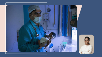
Managing delicate surgical conditions in newborns requires exceptional precision and expertise. Dr. Greeshma Suresh is highly skilled in treating complex congenital anomalies with advanced surgical techniques. Her compassionate approach ensures improved outcomes and safer recovery for neonates.

Minimally Invasive Pediatric Surgery Expertise

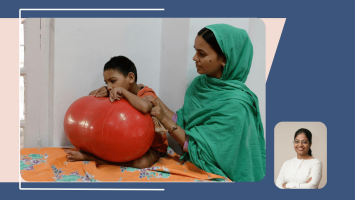
Dr. Greeshma Suresh specializes in minimally invasive pediatric procedures that reduce pain, scarring, and hospital stay. Her expertise ensures faster recovery and enhanced patient comfort.

Comprehensive Anorectal Malformation Management

Early diagnosis and surgical correction are crucial for optimal outcomes in anorectal malformations. Dr. Greeshma Suresh brings extensive experience in performing precise corrective surgeries in children. She focuses on long-term functional outcomes and quality of life.

Specialized TEF Repair & Neonatal Care

Dr. Greeshma Suresh is proficient in managing and surgically correcting TEF with a high success rate. Her expertise ensures safe intervention and improved survival in newborns.

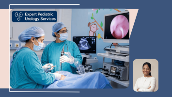
Expert Pediatric Urology Services

Dr. Greeshma Suresh has vast experience in treating pediatric urological conditions such as undescended testes, hypospadias, and hydronephrosis. She delivers patient-centric care with excellent surgical outcomes.

Safe & Effective Pediatric Appendicectomy

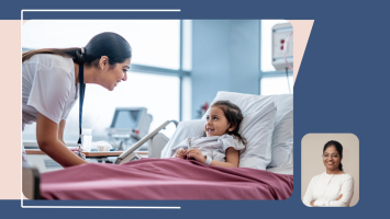
Dr. Greeshma Suresh is skilled in performing safe and effective appendicectomy procedures in pediatric patients. Her approach focuses on timely intervention and quick recovery.

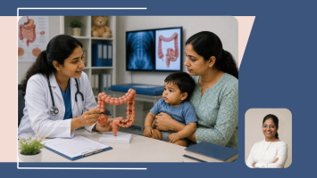
Hirschsprung Disease Treatment

Hirschsprung disease is a congenital condition caused by the absence of nerve cells in a segment of the large bowel, producing functional obstruction, and requires surgical removal of the affected bowel segment with restoration of normal bowel continuity. Dr Greeshma Suresh provides expert diagnosis and surgical management of Hirschsprung disease at Vatsalya Children Hospital, Varanasi, including suction rectal biopsy for confirmation of diagnosis, definitive pull-through surgery to remove the aganglionic segment, and structured long-term follow-up to monitor bowel function and screen for enterocolitis. Her experience in neonatal and paediatric colorectal surgery, combined with her research publications on congenital gastrointestinal conditions, ensures evidence-based management tailored to each child's specific anatomy and clinical condition. Written by Dr Greeshma Suresh, MBBS (NEIGRIHMS), MS General Surgery (Silchar Medical College), MCh Pediatric Surgery (Institute of Medical Sciences, BHU, Varanasi), Paediatric Surgeon, Vatsalya Children Hospital, Mahavir Road, Orderly Bazar, Varanasi, Uttar Pradesh 221002.

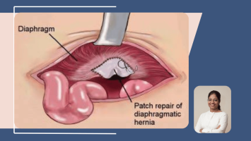
Congenital Diaphragmatic Hernia (CDH) Repair

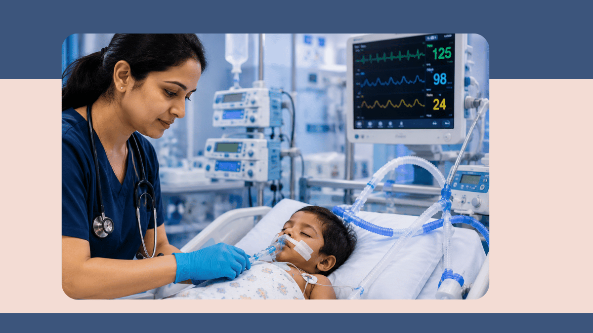
Congenital diaphragmatic hernia (CDH) is a life-threatening neonatal condition in which a defect in the diaphragm allows abdominal organs to herniate into the chest cavity, compressing the developing lungs and producing respiratory failure at birth that requires urgent stabilisation and surgical repair. Dr Greeshma Suresh provides surgical repair of congenital diaphragmatic hernia at Vatsalya Children Hospital, Varanasi, as part of a multidisciplinary management approach involving neonatology, paediatric anaesthesia, and surgical teams. Surgical repair involves returning the herniated organs to the abdomen and closing the diaphragmatic defect, and is performed once the newborn has been stabilised on respiratory support. Her MCh training at BHU's Institute of Medical Sciences and her neonatal surgical experience equip her to manage this technically demanding and time-sensitive condition. Written by Dr Greeshma Suresh, MBBS (NEIGRIHMS), MS General Surgery (Silchar Medical College), MCh Pediatric Surgery (Institute of Medical Sciences, BHU, Varanasi), Paediatric Surgeon, Vatsalya Children Hospital, Mahavir Road, Orderly Bazar, Varanasi, Uttar Pradesh 221002.

PUV Diagnosis: Why Antenatal Detection Changes What Happens to the Kidneys

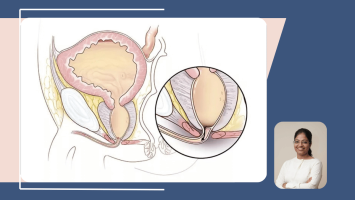
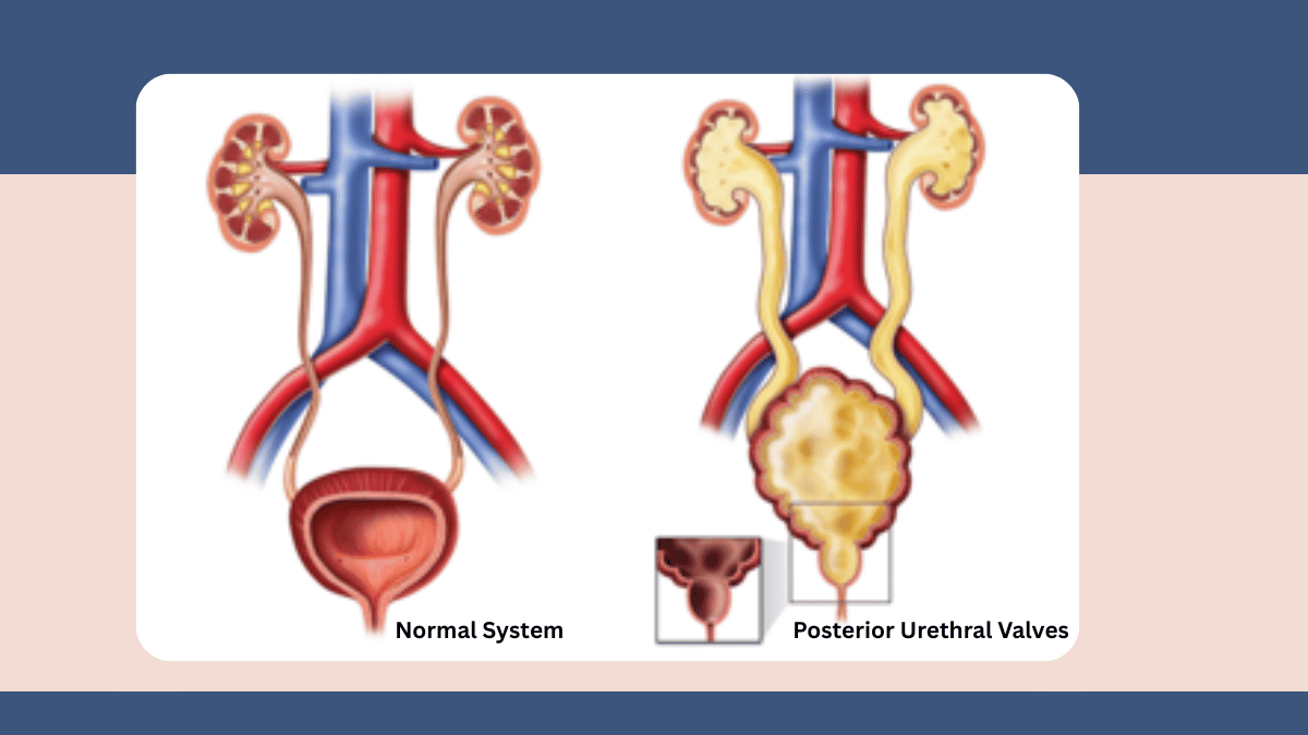
Posterior urethral valves is a condition where the single most powerful determinant of long-term kidney outcome is the timing of diagnosis. The valve obstruction begins causing kidney damage in fetal life, during the critical months of renal development, and every day of untreated obstruction after birth adds to the kidney's burden. Antenatal diagnosis, now achievable through routine second-trimester ultrasound, gives the neonatal and paediatric surgical team the ability to be fully prepared at delivery and to begin treatment within hours of birth. The antenatal ultrasound features that raise the suspicion of PUV in a male fetus include a persistently dilated, thick-walled bladder that does not empty fully between scan images, bilateral hydronephrosis with dilated ureters, and oligohydramnios, a reduced volume of amniotic fluid caused by the fetus producing less urine than normal because the kidney damage has reduced their urinary output. Oligohydramnios in the context of renal obstruction carries its own additional risk to fetal lung development, because the fetal lungs develop by practising breathing movements in amniotic fluid, and severe or prolonged oligohydramnios can result in pulmonary hypoplasia alongside the renal problem. Dr. Greeshma Suresh, MCh Paediatric Surgery from the Institute of Medical Sciences, Banaras Hindu University, where her MCh research specifically investigated aldosterone and renin levels as prognostic biomarkers in PUV, manages PUV at Vatsalya Children Hospital, Varanasi with the combined understanding of the surgical anatomy and the deeper hormonal biology of what obstruction does to the developing kidney. In the delivery room or in the first hours of life, a urethral catheter is placed immediately in any newborn male with known or suspected PUV to drain the bladder acutely and relieve the outflow obstruction. This single intervention begins to reduce the back-pressure on the kidneys before any investigation or definitive treatment takes place. The definitive diagnostic procedure is the micturating cystourethrogram (MCUG), in which contrast dye is instilled into the bladder through the catheter and X-ray images are taken as the baby attempts to void. The MCUG demonstrates the characteristic appearance of a dilated posterior urethra with the impression of the obstructing valve, confirming the diagnosis. Renal ultrasound and blood tests for creatinine and electrolytes are performed simultaneously to establish the baseline of kidney function. The creatinine nadir, the lowest creatinine level reached in the first weeks after birth as the maternal creatinine gradually clears from the baby's bloodstream, is one of the most clinically important values in PUV management: it provides the best early estimate of the baby's own underlying kidney function, and it predicts long-term renal outcome better than the birth creatinine level alone.

Endoscopic Valve Ablation and Long-Term Follow-Up: The Surgery and What Comes After

Endoscopic valve ablation is the definitive surgical treatment for posterior urethral valves, and in newborns who are large enough for the cystoscope to pass safely through the urethra, it is the treatment of choice from the earliest days of life. The procedure requires no skin incision. A small cystoscope is passed through the urethra into the bladder under general anaesthesia, and the abnormal obstructing tissue of the posterior urethral valve is divided using a fine diathermy hook or cold knife passed through the instrument channel of the cystoscope. The division of the valve tissue immediately removes the obstruction to urinary outflow. The procedure takes approximately 20 to 30 minutes. A urethral catheter remains in place for one to two days after the ablation to allow the urethra to rest, and is then removed. The technical challenge of PUV ablation in the newborn period is the size of the urethra: it is a narrow structure in a newborn male, and the cystoscope must be small enough to pass through without damaging the urethral mucosa. In very small or premature newborns whose urethra is too narrow to accommodate even the smallest available paediatric cystoscope safely, the immediate endoscopic ablation is deferred. A temporary vesicostomy is performed instead: a small opening is created between the bladder and the skin of the lower abdomen, through which urine drains freely without having to pass through the obstructed urethra. The vesicostomy protects the kidneys from ongoing back-pressure while the baby grows to a size at which endoscopic ablation can be performed safely. Once ablation is complete, whether immediately or following a period with a vesicostomy, the urinary tracts are monitored with serial renal ultrasound scans to assess how the hydronephrosis resolves and how the bladder wall thickness changes. Kidney function is monitored regularly with blood tests. Dr. Greeshma Suresh is clear that parents need to understand the important distinction between what ablation achieves and what it cannot achieve. Endoscopic valve ablation removes the obstruction. It cannot reverse the developmental damage that the kidneys and bladder sustained during the months of fetal obstruction. Many boys with PUV have some degree of bladder dysfunction after successful ablation, because the bladder has spent months developing against a high-resistance obstruction and has acquired patterns of abnormal storage, reduced compliance, or incomplete emptying that do not automatically normalise when the obstruction is removed. Bladder function assessment, bladder training, anticholinergic medication, or intermittent catheterisation may be part of the long-term management plan. Some boys have significant residual renal impairment that requires ongoing nephrological care. The MCh research into aldosterone and renin as biomarkers of kidney hormonal adaptation in PUV informs the understanding of why some kidneys recover more function after ablation than others, and why continued surveillance of these hormonal markers alongside standard renal function tests provides a more complete picture of kidney health over time. At Vatsalya Children Hospital, Varanasi, every boy treated for PUV is enrolled in a structured long-term follow-up programme involving serial renal imaging, kidney function testing, bladder function assessment, and blood pressure monitoring, because the outcomes in this condition are determined as much by the quality of ongoing surveillance as by the quality of the initial surgery.

Dr. Greeshma Suresh 's Expert Tips & Health Insights

Latest

Articles by Dr. Greeshma Suresh

https://d2vvc091k8tmib.cloudfront.net/s3fs-public/2026-09/Posterior%20Urethral%20Valves%20in%20Male%20Newborns.png
https://d2vvc091k8tmib.cloudfront.net/s3fs-public/2026-04/Dr.%20Baby%20Sana%20K%20%2811%29.jpg
By: Dr. Greeshma Suresh
04 September 2026

Posterior Urethral Valves in Male Newborns: What Parents Need to Know

A Paediatric Surgeon whose own published research covers posterior urethral valves explains what PUV is, why it causes kidney damage before birth, how it is diagnosed and treated, and what the long-term kidney outlook is for affected boys, at Vatsalya Children Hospital, Varanasi.
Read More
https://d2vvc091k8tmib.cloudfront.net/s3fs-public/2026-08/Hirschsprung%20Disease%20in%20Newborns%20What%20It%20Is%2C%20How%20It%20Is%20Diagnosed%2C%20and%20What%20Surgery%20Achieves.png
https://d2vvc091k8tmib.cloudfront.net/s3fs-public/2026-04/Dr.%20Baby%20Sana%20K%20%2811%29.jpg
By: Dr. Greeshma Suresh
05 August 2026

Hirschsprung Disease in Newborns: What It Is, How It Is Diagnosed, and…

Hirschsprung disease causes bowel obstruction in newborns due to missing nerve cells - a paediatric surgeon explains diagnosis, pull-through surgery, and the long-term risk of enterocolitis.
Read More
https://d2vvc091k8tmib.cloudfront.net/s3fs-public/2026-07/Neonatal%20Surgery_%20A%20Complete%20Guide%20for%20Parents%20Facing%20a%20Newborn%20Surgical%20Diagnosis.png
https://d2vvc091k8tmib.cloudfront.net/s3fs-public/2026-04/Dr.%20Baby%20Sana%20K%20%2811%29.jpg
By: Dr. Greeshma Suresh
02 July 2026

Neonatal Surgery: A Complete Guide for Parents Facing a Newborn Surgic…

Learn about neonatal surgery, common newborn surgical conditions, NICU care, recovery, and treatment options with expert guidance from Dr. Greeshma Suresh, Varanasi.
Read More
https://d2vvc091k8tmib.cloudfront.net/s3fs-public/2026-07/9_0.png
https://d2vvc091k8tmib.cloudfront.net/s3fs-public/2026-04/Dr.%20Baby%20Sana%20K%20%2811%29.jpg
By: Dr. Greeshma Suresh
31 March 2026

Appendicitis in Children: Early Warning Signs and Emergency Treatment …

Is your child having sudden stomach pain, fever & vomiting? These could be early signs of appendicitis. Dr. Greeshma Suresh, pediatric surgeon in Varanasi, explains symptoms & emergency treatment.
Read More
https://d2vvc091k8tmib.cloudfront.net/s3fs-public/2026-07/10.png
https://d2vvc091k8tmib.cloudfront.net/s3fs-public/2026-04/Dr.%20Baby%20Sana%20K%20%2811%29.jpg
By: Dr. Greeshma Suresh
31 March 2026

Undescended Testes in Children: Causes, Diagnosis, and Surgical Manage…

Undescended testes in children needs early surgery for best results. Dr. Greeshma Suresh, pediatric surgeon in Varanasi, explains causes, cryptorchidism diagnosis & orchiopexy treatment options.
Read More
https://d2vvc091k8tmib.cloudfront.net/s3fs-public/2026-07/11.png
https://d2vvc091k8tmib.cloudfront.net/s3fs-public/2026-04/Dr.%20Baby%20Sana%20K%20%2811%29.jpg
By: Dr. Greeshma Suresh
31 March 2026

Pediatric Hernia: Signs, Types, and When Surgery is Needed in Children

Noticed a bulge in your child's groin or belly? It could be a hernia. Dr. Greeshma Suresh, pediatric surgeon in Varanasi, explains hernia types, warning signs & when surgery is needed in children.
Read More
https://d2vvc091k8tmib.cloudfront.net/s3fs-public/2026-07/12.png
https://d2vvc091k8tmib.cloudfront.net/s3fs-public/2026-04/Dr.%20Baby%20Sana%20K%20%2811%29.jpg
By: Dr. Greeshma Suresh
31 March 2026

Common Pediatric Surgical Conditions in Children: Symptoms, Causes, an…

Worried about your child needing surgery? Dr. Greeshma Suresh, pediatric surgeon in Varanasi, covers common surgical conditions in children — symptoms, causes & the right time to consult a surgeon.
Read More
https://d2vvc091k8tmib.cloudfront.net/s3fs-public/2026-07/13.png
https://d2vvc091k8tmib.cloudfront.net/s3fs-public/2026-04/Dr.%20Baby%20Sana%20K%20%2811%29.jpg
By: Dr. Greeshma Suresh
31 March 2026

Minimally Invasive Pediatric Surgery: Benefits, Procedures, and Recove…

Laparoscopic surgery offers children faster recovery, less pain & minimal scarring. Dr. Greeshma Suresh, pediatric surgeon in Varanasi, explains minimally invasive surgery benefits & procedures.
Read More
https://d2vvc091k8tmib.cloudfront.net/s3fs-public/2026-07/14.png
https://d2vvc091k8tmib.cloudfront.net/s3fs-public/2026-04/Dr.%20Baby%20Sana%20K%20%2811%29.jpg
By: Dr. Greeshma Suresh
31 March 2026

Congenital Anomalies in Newborns: Early Diagnosis and Pediatric Surgic…

Congenital anomalies in newborns, commonly known as birth defects in babies, are structural or functional abnormalities present at birth. These congenital disorders in newborns can affect various organs and systems, including congenital heart defects in newborns, neural tube defects in babies, cleft lip and palate in newborns, and other congenital malformations in infants.
Read More

Dr. Greeshma Suresh 's Patient Testimonial Reviews

Leave a Review

Patient Reviews

Dr. Greeshma Suresh

Leave a Review

Frequently Asked Questions

Frequently Asked Questions

View all

My son has been successfully treated for PUV. How long does he need to be followed up?

The direct answer is: lifelong. And I want to explain precisely why this is not an overstatement or an excess of caution, because understanding the reasons for long-term follow-up in PUV changes how families engage with it. Posterior urethral valves is not a condition where the definitive surgery produces a cure in the sense that the underlying biology returns to normal. Endoscopic valve ablation removes the obstruction. It does not reverse the developmental changes that the kidneys and bladder sustained during the months of fetal obstruction. The bladder that spent the entire fetal period developing against a high-resistance outflow obstruction has acquired structural and functional characteristics that persist after the valve is gone: increased wall thickness, reduced compliance (less ability to stretch and store urine at low pressure), abnormal patterns of emptying, and in some boys a poorly compliant bladder that generates high pressures during filling and continues to damage the kidneys from below even after the anatomical obstruction above has been removed. Identifying and managing this bladder dysfunction is one of the most important aspects of long-term PUV care, because a high-pressure bladder after ablation can cause ongoing kidney damage by the same back-pressure mechanism as the original valve. Regular bladder function assessment, which may include urodynamic testing, is therefore part of the follow-up schedule. The kidney function monitoring component of follow-up involves regular blood tests for creatinine, electrolytes, and markers of kidney tubular function, which assess how the kidneys are performing over time, and whether function is stable, improving, or declining. Blood pressure monitoring is equally important: high blood pressure is both more common in boys with PUV and damaging to kidneys that already have reduced reserve capacity. As these boys grow through childhood, adolescence, and into adulthood, the follow-up transitions from paediatric surgical and nephrological care to adult urology and nephrology. At Vatsalya Children Hospital, Varanasi, the structured long-term follow-up programme for PUV patients is not a precaution: it is the clinical infrastructure that translates a good initial surgical outcome into the best possible long-term kidney and bladder health for every boy we treat.

Related reading: 

Posterior Urethral Valves in Male Newborns: What Parents Need to Know
Neonatal Surgery: A Complete Guide for Parents Facing a Newborn Surgical Diagnosis
Hirschsprung Disease in Newborns: What It Is, How It Is Diagnosed, and What Surgery Achieves
Congenital Anomalies in Newborns: Early Diagnosis and Pediatric Surgical Treatment Options


Written by Dr. Greeshma Suresh, MBBS, MS General Surgery, MCh Paediatric Surgery (Institute of Medical Sciences, Banaras Hindu University), Paediatric Surgeon, Vatsalya Children Hospital, Varanasi 221002. Phone: +91 9838585111. 

Will my son's kidneys recover after the valve is removed?

This is the question that matters most to parents of a child with PUV, and I want to answer it honestly. Some recovery of kidney function after valve ablation is common, particularly in the first months after the obstruction is relieved. The kidneys that have been working under pressure are no longer subjected to back-pressure once the valve is ablated, and the hydronephrosis typically begins to reduce on serial ultrasound imaging. Blood creatinine levels may improve in the first months after ablation. But the honest clinical picture is more complex than simple recovery. The kidneys of a baby with PUV have been developing under obstruction since the fetal period: the back-pressure began before the kidneys were fully formed, during the critical developmental window in the second and third trimesters of pregnancy when kidney development depends on unobstructed urine flow and normal amniotic fluid dynamics. Kidney tissue that was damaged or failed to develop normally during this fetal period cannot be regenerated by removing the valve after birth. What the creatinine nadir, the lowest creatinine level in the first weeks of life, tells us is crucial: a creatinine nadir below approximately 80 micromoles per litre generally indicates that enough functioning kidney tissue developed to support long-term kidney health. A nadir that remains persistently elevated indicates more significant underlying renal impairment. My MCh research on aldosterone and renin levels in PUV patients at IMS BHU contributes to the understanding of how the kidney's hormonal systems adapt to obstruction, and these biomarkers alongside standard function tests give a more nuanced picture of renal health over time than creatinine alone. Some boys with PUV go on to have normal kidney function with structured follow-up and no long-term impairment. Others have persistent reduction in kidney function that requires ongoing nephrological care, blood pressure management, and in some eventually the consideration of renal replacement therapy. The honest answer is that I cannot tell every family at birth exactly where on this spectrum their son will fall, but I can tell them that the combination of early diagnosis, prompt treatment, careful long-term surveillance, and expert management of bladder function gives their son's kidneys the best possible chance of the best possible outcome.

Related reading: 

Posterior Urethral Valves in Male Newborns: What Parents Need to Know
Neonatal Surgery: A Complete Guide for Parents Facing a Newborn Surgical Diagnosis
Hirschsprung Disease in Newborns: What It Is, How It Is Diagnosed, and What Surgery Achieves
Congenital Anomalies in Newborns: Early Diagnosis and Pediatric Surgical Treatment Options


Written by Dr. Greeshma Suresh, MBBS, MS General Surgery, MCh Paediatric Surgery (Institute of Medical Sciences, Banaras Hindu University), Paediatric Surgeon, Vatsalya Children Hospital, Varanasi 221002. Phone: +91 9838585111. 

The surgeon said my newborn is too small for the cystoscope. What does vesicostomy mean and is it safe?

Vesicostomy is a temporary surgical procedure in which a small opening is created between the bladder and the surface of the skin of the lower abdomen, allowing urine to drain freely from the bladder without having to pass through the urethra at all. In a baby with posterior urethral valves whose urethra is too narrow to allow the cystoscope to pass safely, the vesicostomy is the alternative that provides the same essential benefit as immediate endoscopic ablation: it removes the back-pressure on the kidneys by giving the urine a route out of the bladder that bypasses the obstruction entirely. The urethra of a very small or premature newborn male is an extremely narrow structure. The smallest paediatric cystoscopes are designed to pass safely in newborns of sufficient size, but attempting to pass even the smallest instrument through a urethra that is not ready for it risks urethral injury, scarring, and stricture formation, which would create a new and permanent obstruction on top of the one being treated. The safer clinical decision in these situations is the vesicostomy, which protects the kidneys immediately without the risk of urethral injury. The vesicostomy is a small surgical operation under general anaesthesia that takes approximately 30 to 45 minutes. The opening created is deliberate and controlled: it is not a wound or a complication, but a planned diversion. Urine drains through this opening into a nappy in the usual way, and the baby is otherwise managed normally. As the baby grows and the urethra reaches the diameter that allows the cystoscope to pass safely, endoscopic valve ablation is performed, and after the ablation has healed and the urinary flow through the urethra is confirmed to be normal, the vesicostomy is surgically closed. The entire sequence is planned from the beginning: the vesicostomy is not a failure to do the right operation, it is the right operation for this specific baby at this specific size. At Vatsalya Children Hospital, Varanasi, the decision between immediate endoscopic ablation and temporary vesicostomy is made on the individual baby's size, urethra calibre, and overall clinical stability, with the single goal of protecting the kidneys from further damage in the safest way available for that particular baby.


Written by
Dr. Greeshma Suresh, MBBS, MS General Surgery, MCh Paediatric Surgery (Institute of Medical Sciences, Banaras Hindu University), Paediatric Surgeon, Vatsalya Children Hospital, Varanasi 221002. Phone: +91 9838585111. 

My son's antenatal scan showed a dilated bladder and enlarged kidneys. What should happen next?

An antenatal scan finding of a dilated, thick-walled bladder that does not empty, combined with bilateral enlarged kidneys (bilateral hydronephrosis) and dilated ureters in a male fetus, is a pattern that should raise immediate suspicion of posterior urethral valves and should prompt specialist referral without delay. This is not a finding that should be monitored with repeated antenatal scans alone and reassessed only after birth: the earlier the diagnosis is confirmed and the clinical team is prepared, the better the outcome for the baby's kidneys. The steps that should follow this antenatal finding are: a detailed fetal anomaly scan by a specialist sonologist to characterise the urinary tract findings more precisely and to assess the volume of amniotic fluid, as oligohydramnios (reduced amniotic fluid) indicates that the kidney damage has already reduced fetal urine output significantly; a referral to a fetal medicine or paediatric surgery specialist for counselling and birth planning; planning for the delivery to take place at a hospital where a paediatric surgeon and neonatal team are available, or where postnatal transfer to such a hospital is immediately available; and pre-birth discussion with the family about what will happen in the first hours of the baby's life. In the delivery room, a urethral catheter will be placed in the baby immediately or in the first hours of life to drain the bladder and relieve the obstruction acutely. Investigation of the urinary tract and assessment of kidney function begins within the first day. The confirmation diagnostic procedure, the MCUG, is performed as soon as the baby is stable enough. The single most important message I give to parents receiving this antenatal finding is: seek a specialist paediatric surgical review now, plan the birth at or near a centre with paediatric surgical capability, and ensure the team is ready at delivery. The time between this scan and delivery is not waiting time. It is preparation time, and that preparation changes outcomes.

Related reading: 

Posterior Urethral Valves in Male Newborns: What Parents Need to Know
Neonatal Surgery: A Complete Guide for Parents Facing a Newborn Surgical Diagnosis
Hirschsprung Disease in Newborns: What It Is, How It Is Diagnosed, and What Surgery Achieves
Congenital Anomalies in Newborns: Early Diagnosis and Pediatric Surgical Treatment Options
Written by Dr. Greeshma Suresh, MBBS, MS General Surgery, MCh Paediatric Surgery (Institute of Medical Sciences, Banaras Hindu University), Paediatric Surgeon, Vatsalya Children Hospital, Varanasi 221002. Phone: +91 9838585111. 

Can a baby who has had neonatal surgery feed normally afterwards?

Can a baby who has had neonatal surgery feed normally afterwards?

Link copied to clipboard!

Reach Dr. Greeshma Suresh

Vatsalya Children Hospital

Mahavir Road, near Mahavir Mandir Crossing, Orderly Bazar, Varanasi, Uttar Pradesh 221002