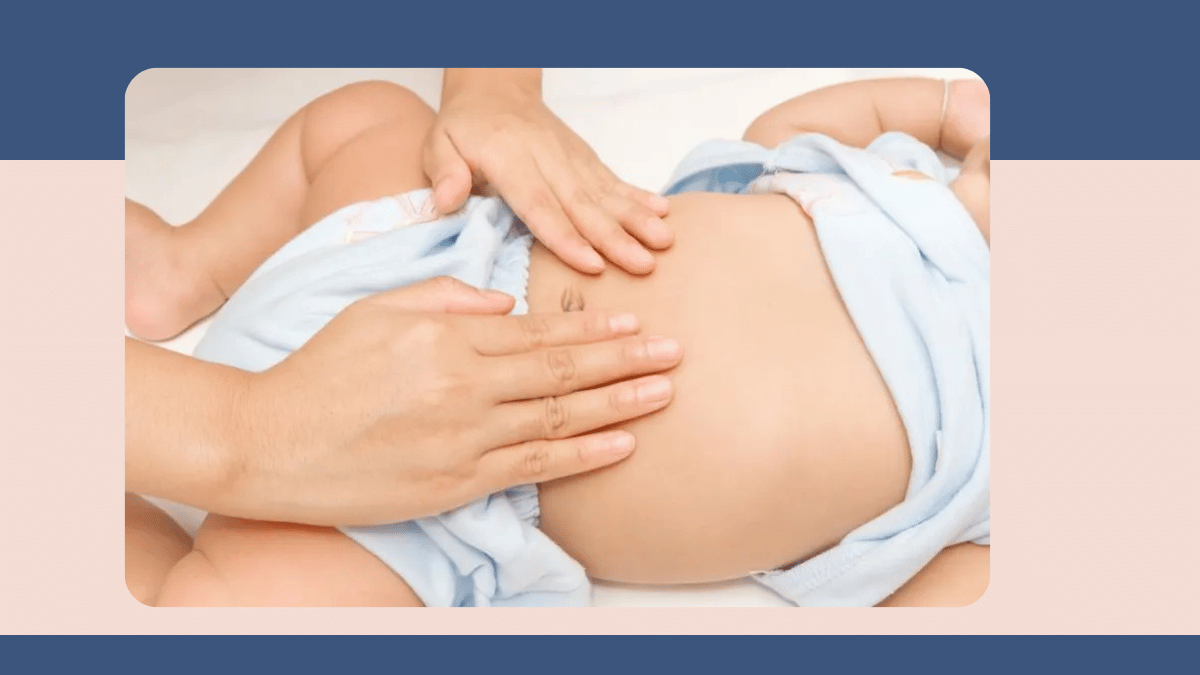
One of the conditions I see regularly as a paediatric surgeon at Vatsalya Children Hospital, Varanasi, is Hirschsprung disease - a congenital abnormality of the bowel that typically presents in the newborn period but can be missed for months or years when its presentation is less acute. It is a condition that most parents have never heard of before their child's diagnosis, and yet it accounts for a significant proportion of neonatal surgical admissions. Understanding what Hirschsprung disease is, why it causes the symptoms it does, and what surgery achieves helps families approach a difficult situation with a clearer picture of what to expect from treatment and follow-up.
What Hirschsprung disease is and why it causes bowel obstruction
During normal foetal development, nerve cells called ganglion cells migrate along the bowel from top to bottom, providing the neural infrastructure that allows the bowel to contract and relax in the coordinated wave of movement called peristalsis. In Hirschsprung disease, this migration is incomplete. Ganglion cells are absent from the lowest segment of the large bowel, and in some cases from a longer segment. The aganglionic segment cannot relax, creating a functional obstruction - stool backs up behind the non-relaxing segment, the bowel above it dilates, and the patient develops constipation or complete bowel obstruction. In the newborn period, the most characteristic sign is failure to pass meconium in the first 48 hours of life. Healthy newborns almost always pass meconium, the dark tarry first stool, within 24 to 48 hours. When this does not happen, and the abdomen begins to distend and the baby vomits, Hirschsprung disease is one of the first diagnoses to consider. In older infants and children with a milder or shorter segment of involvement, the condition may present as severe chronic constipation that has not responded to standard treatment.
Diagnosis: why the suction rectal biopsy is the gold standard
The definitive diagnosis of Hirschsprung disease requires a tissue sample from the bowel wall to confirm the absence of ganglion cells. This is obtained through a suction rectal biopsy, a procedure that collects a small sample of the rectal mucosa and submucosa without requiring a general anaesthetic or an incision. The sample is examined by a pathologist for the presence of ganglion cells and for the enzyme histochemical changes associated with Hirschsprung disease. The suction rectal biopsy is performed at Vatsalya Children Hospital as part of the standard diagnostic work-up for any newborn or infant suspected of having Hirschsprung disease. Contrast enema and plain abdominal X-ray provide supporting information and help define the level of the aganglionic segment, but the biopsy result is what confirms the diagnosis and determines the surgical plan.
Surgical treatment for Hirschsprung disease involves removing the aganglionic segment of bowel and bringing the normally innervated bowel down to the anus in what is called a pull-through procedure. This can be performed in a single stage or with a temporary stoma depending on the baby's condition, the extent of the disease, and the degree of bowel dilation present. Minimally invasive approaches are used for appropriate cases. The goal of surgery is to remove the obstruction, restore normal bowel function, and give the child the best possible chance of achieving long-term bowel control.
Long-term follow-up and the risk of enterocolitis
Surgery for Hirschsprung disease is not the end of the journey. The most important concern in long-term follow-up is Hirschsprung-associated enterocolitis, a potentially serious inflammatory condition of the bowel that can occur both before surgery and in the months and years after the pull-through procedure. Enterocolitis presents with explosive diarrhoea, fever, abdominal distension, and a child who is unwell. It can progress rapidly to a severe illness if not treated promptly, which is why families need to know the warning signs and have clear instructions about when to seek emergency assessment.
Long-term bowel function after pull-through surgery varies. Many children achieve good continence and quality of life, particularly when the aganglionic segment is short. Children with longer segment disease or those with associated conditions may need ongoing bowel management support. Regular follow-up with a paediatric surgeon who knows the specific anatomy of each case and the child's bowel function history is essential, not just in the first year after surgery but throughout childhood. At Vatsalya Children Hospital, I structure post-operative follow-up to monitor bowel function, identify signs of enterocolitis early, and adjust management as the child grows.
To book a consultation with Dr. Greeshma Suresh at Vatsalya Children Hospital, Mahavir Road, Orderly Bazar, Varanasi, call +91 9838585111.
Written by Dr. Greeshma Suresh, MBBS, MS (General Surgery), MCh (Pediatric Surgery), Institute of Medical Sciences, Banaras Hindu University, Paediatric Surgeon, Vatsalya Children Hospital, Mahavir Road, Orderly Bazar, Varanasi, Uttar Pradesh 221002. Phone: +91 9838585111.
Related reading
Neonatal Surgery: A Complete Guide for Parents Facing a Newborn Surgical Diagnosis
Congenital Anomalies in Newborns: Early Diagnosis and Pediatric Surgical Treatment Options
Minimally Invasive Pediatric Surgery: Benefits, Procedures, and Recovery Explained
Common Pediatric Surgical Conditions in Children: Symptoms, Causes, and Treatment Guide


Add a Comment