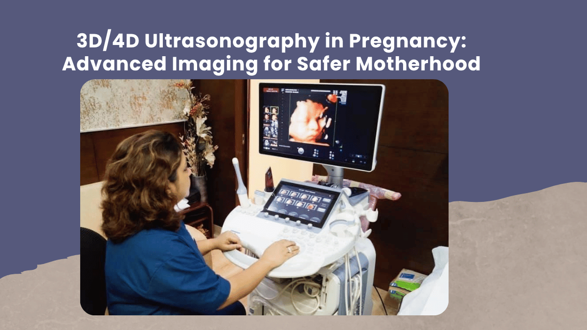
3D/4D ultrasonography has revolutionised modern prenatal care by offering detailed and lifelike images of the developing baby. Unlike traditional 2D ultrasound, 3D ultrasonography provides three-dimensional still images, while 4D ultrasonography adds real-time motion, allowing doctors and parents to observe fetal movements, facial expressions, and structural development more clearly. This advanced prenatal imaging technology improves early detection of congenital anomalies, facial defects, limb abnormalities, and spinal conditions. It also enhances fetal growth monitoring, placental assessment, and evaluation of amniotic fluid levels. For high-risk pregnancies, 3D/4D ultrasound plays a vital role in accurate diagnosis and timely medical intervention, contributing significantly to improved maternal and fetal outcomes.
In my clinical experience, I have seen how 3D/4D ultrasonography not only improves diagnostic accuracy but also reassures expecting parents. Many mothers walk into the scan room feeling anxious, especially if they have had previous complications or are managing a high-risk pregnancy. When they are able to clearly see their baby’s features and movements, it often brings immense emotional comfort. Beyond the emotional connection, I rely on advanced imaging to carefully evaluate fetal anatomy, detect subtle abnormalities, and plan appropriate management well in advance. Early identification allows us to take proactive steps, ensuring safer pregnancy progression and better birth outcomes.
From a medical perspective, 3D/4D ultrasound technology is particularly beneficial in evaluating facial clefts, neural tube defects, skeletal abnormalities, and uterine anomalies. It also assists in assessing fetal behaviour patterns and overall well-being. While these scans provide enhanced clarity, they are recommended based on clinical indications and medical necessity. Responsible use ensures both safety and effectiveness in prenatal diagnosis. Combined with routine antenatal check-ups, blood investigations, and Doppler studies, 3D/4D ultrasonography strengthens comprehensive pregnancy monitoring.
Under the expert guidance of Dr. Khyati Sunildatt Daru, advanced prenatal imaging is integrated into a structured, evidence-based antenatal care plan. Her approach focuses on precision, early risk detection, and personalised pregnancy management. By combining medical expertise with compassionate counselling, she ensures that every mother receives accurate information and confident guidance throughout her pregnancy journey.
Conclusion
3D/4D ultrasonography has become an essential component of modern obstetric care, offering both advanced diagnostic capability and emotional reassurance for families. When performed by a trained specialist and used appropriately, it supports early detection of complications, enhances pregnancy monitoring, and contributes to safer deliveries. With the right medical supervision and timely imaging, expecting parents can move forward with greater confidence, knowing that their baby’s health is being closely and carefully monitored.



Add a Comment