A brain aneurysm diagnosis changes everything about how a person understands their own body. The knowledge that there is a bulge in an artery supplying the brain, one that could rupture without warning, is frightening in a way that is difficult to convey to anyone who has not received it. In my practice as a neurosurgeon and neuroendovascular specialist at SPARSH Hospital, Infantry Road, Bengaluru, managing cerebrovascular conditions including aneurysms, arteriovenous malformations, and carotid artery disease is the work I find most technically demanding and most clinically consequential. I trained in neurosurgery at GB Pant Hospital and Maulana Azad Medical College, New Delhi, one of India's foremost neurosurgical training institutions, and cerebrovascular surgery has been a central part of my practice throughout my career. This guide is written for every patient and family trying to understand what a brain aneurysm is, what it means for their health, and what the treatment options actually involve.
What is a brain aneurysm?
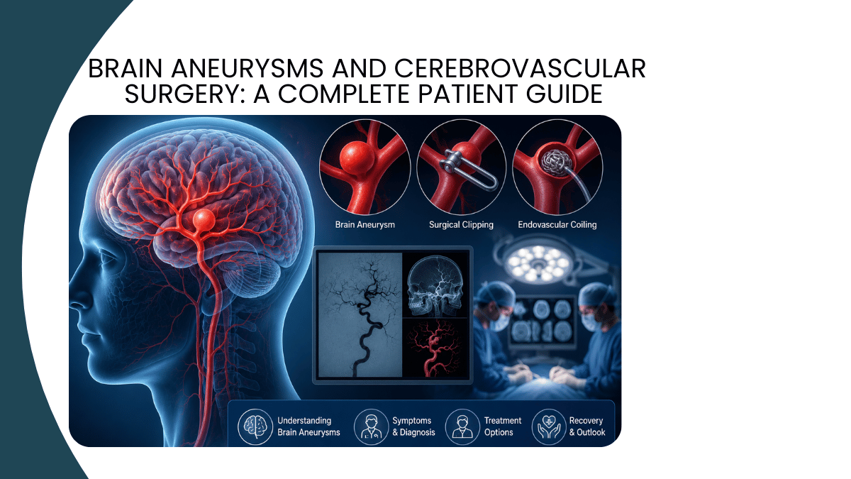
A brain aneurysm is a localised weakening and ballooning of an arterial wall within or near the brain. The aneurysm forms at a point where the arterial wall is structurally weak, most commonly at the junction between two arteries, and gradually expands under the pressure of blood flow. Most aneurysms are saccular, or berry-shaped, protruding from the side of an artery like a small blister. Fusiform aneurysms, which involve a diffuse widening of an entire arterial segment rather than a localised bulge, are less common and present different treatment challenges. The critical risk associated with any intracranial aneurysm is rupture: when the aneurysmal wall fails, blood escapes under arterial pressure into the subarachnoid space surrounding the brain, producing a subarachnoid haemorrhage that is immediately life-threatening and carries a very high risk of permanent neurological injury or death even with prompt treatment.
How common are brain aneurysms and who is at risk?
Population studies suggest that intracranial aneurysms are present in approximately 2 to 3 percent of the general population, meaning that millions of people carry an unruptured aneurysm without knowing it. The majority of these aneurysms never rupture and are discovered incidentally on imaging obtained for another reason. However, certain factors increase both the likelihood of having an aneurysm and the risk of rupture. Risk factors for brain aneurysm rupture include hypertension, cigarette smoking, heavy alcohol use, a family history of intracranial aneurysms particularly in first-degree relatives, certain connective tissue disorders including Ehlers-Danlos syndrome type IV and Marfan syndrome, polycystic kidney disease, and previous aneurysmal subarachnoid haemorrhage. Aneurysm size and morphology also influence rupture risk: larger aneurysms and those with an irregular shape or daughter sac are more likely to rupture than small, smooth-walled lesions.
How aneurysms are discovered
Unruptured aneurysms are discovered in three main ways. Incidental discovery occurs when an aneurysm is identified on brain imaging, typically an MRI or CT scan, performed for an unrelated indication such as headache evaluation or screening. Family screening occurs in individuals with a known first-degree relative who has had an aneurysmal subarachnoid haemorrhage: the risk of harbouring an aneurysm is approximately three to five times higher in this group, and screening with MR angiography is warranted. Symptomatic discovery occurs when an unruptured aneurysm causes symptoms through compression of adjacent structures: a large posterior communicating artery aneurysm compressing the oculomotor nerve, for example, may present as a sudden painful third nerve palsy with ptosis and a dilated pupil, a presentation I treat as a neurosurgical emergency warranting same-day imaging and evaluation.
The discovery of a ruptured aneurysm is almost always dramatic. Subarachnoid haemorrhage produces the classic thunderclap headache, described by patients as the worst headache of their life, of sudden explosive onset unlike any previous headache, often accompanied by nausea, vomiting, photophobia, neck stiffness, and in severe cases, loss of consciousness. This presentation warrants emergency medical attention and immediate CT imaging of the brain, which detects subarachnoid haemorrhage with high sensitivity in the first 24 hours. When clinical suspicion remains high despite a normal CT, lumbar puncture is performed to detect xanthochromia, the yellow discolouration of the cerebrospinal fluid that results from haemoglobin breakdown.
How are brain aneurysms diagnosed and evaluated?
Once an aneurysm is suspected or identified on non-invasive imaging, a comprehensive vascular evaluation is required to define its size, shape, location, relationship to adjacent arteries, and the anatomy of the surrounding vasculature before any treatment decision is made. CT angiography provides rapid high-resolution three-dimensional images of the intracranial vasculature and is the first-line investigation for both suspected subarachnoid haemorrhage and incidentally discovered vascular lesions. MR angiography avoids radiation and is preferred for screening in lower-risk settings and for follow-up imaging. Digital subtraction angiography (DSA), the gold standard for cerebrovascular imaging, uses catheter-based contrast injection under fluoroscopic guidance to produce detailed images of the aneurysm and its parent artery, including information about blood flow dynamics that non-invasive imaging cannot capture. I perform cerebral angiography as part of my neuroendovascular practice, and DSA is an essential part of the pre-treatment evaluation for any patient being considered for endovascular intervention.
The two treatment approaches: surgical clipping versus endovascular coiling
When treatment is indicated for a brain aneurysm, there are two fundamentally different approaches available: microsurgical clipping and endovascular coiling. Both aim to exclude the aneurysm from the circulation and eliminate the risk of rupture, but they achieve this through completely different mechanisms, through different anatomical routes, and with different risk profiles for different patients. The decision between clipping and coiling is one of the most important clinical judgements in cerebrovascular surgery, and it requires a neurosurgeon with genuine competence in both approaches to evaluate each case without bias towards one technique.
Microsurgical clipping
Microsurgical clipping involves opening the skull through a craniotomy, navigating through the brain's natural anatomical corridors using a high-powered operating microscope, and placing a titanium clip across the neck of the aneurysm to exclude it permanently from the circulation. The clip is permanent, made of titanium, and MRI-compatible. When successfully placed across the aneurysm neck, it provides immediate and definitive aneurysm obliteration. Brain aneurysm clipping is particularly effective for aneurysms with a broad neck that would not be securely occluded by coiling, aneurysms in locations more safely approached surgically, and in cases where a clot compressing the brain needs to be evacuated at the same time as the aneurysm is secured. The surgical approach is demanding, requires microsurgical experience and familiarity with the relevant anatomy, and carries specific risks including stroke from parent artery injury and cranial nerve injury from retraction.
Endovascular coiling and flow diversion
Endovascular treatment accesses the aneurysm from inside the vascular system rather than through a craniotomy. A catheter is introduced through a small puncture in the groin artery and navigated under fluoroscopic guidance through the aorta, carotid arteries, and intracranial vessels to the aneurysm itself. Platinum coils are then deployed through the catheter into the aneurysm sac, packing it to promote clot formation and isolate it from the circulation. For wide-necked aneurysms that cannot be coiled alone, a stent is first deployed across the aneurysm neck to provide a scaffold supporting the coils, or a flow diverter, a high-porosity mesh stent, is placed across the aneurysm neck to redirect blood flow away from the aneurysm and induce progressive thrombosis. I perform the full range of endovascular aneurysm coiling procedures including simple coiling, balloon-assisted coiling, stent-assisted coiling, and flow diversion at SPARSH Hospital. The advantage of endovascular treatment is the avoidance of craniotomy: patients recover faster, the procedure can be performed under general anaesthesia or conscious sedation, and access to deeply situated aneurysms that would require complex surgical approaches is achieved through the same catheter-based route regardless of location.
How the choice is made
The landmark ISAT trial demonstrated that endovascular coiling produces better clinical outcomes than surgical clipping for ruptured aneurysms suitable for both treatments, with fewer patients dead or dependent at one year in the coiling group. This evidence has shifted practice significantly towards endovascular treatment for most ruptured aneurysms. However, the ISAT result applies to patients whose aneurysms were suitable for both techniques: the many patients with aneurysms that are better suited to clipping due to size, morphology, or location still require surgical intervention. My practice is to offer each patient the treatment that is most appropriate for their specific aneurysm, based on its anatomy, and to have the technical capability to perform either procedure. A cerebrovascular surgeon who can only offer one technique is not in a position to provide this kind of individualised recommendation.
Arteriovenous malformations: a different cerebrovascular challenge
An arteriovenous malformation (AVM) is an abnormal tangle of blood vessels connecting arteries directly to veins without the intervening capillary bed that normally exists between them. AVMs are congenital lesions that present most commonly in young adults with haemorrhage, seizures, headaches, or progressive neurological deficit. The annual risk of haemorrhage from an untreated AVM is approximately 2 to 4 percent, which over a lifetime produces a substantial cumulative risk, particularly in younger patients.
The management of AVMs is one of the most complex decision-making processes in cerebrovascular surgery. Treatment options include microsurgical resection, stereotactic radiosurgery, AVM embolisation using liquid embolic agents to reduce blood flow through the malformation prior to surgery or as the primary treatment in selected cases, and in many cases a combination of all three modalities. I perform AVM embolisation as part of my neuroendovascular practice, working in collaboration with radiosurgery colleagues where combined treatment is indicated. The decision about whether and how to treat an AVM requires careful weighing of the lesion's natural history risk against the treatment risk, which varies substantially with the AVM's size, location, and venous drainage pattern.
Carotid artery disease and carotid artery stenting
Atherosclerotic disease of the carotid arteries, the main arteries supplying blood to the brain from the neck, is one of the most important preventable causes of ischaemic stroke. Carotid artery stenosis narrows the arterial lumen and creates a source of embolic material that can travel to the brain and cause stroke or TIA. When stenosis is significant in a patient who has had a stroke or TIA attributable to the carotid lesion, or when stenosis is very severe even in an asymptomatic patient, revascularisation reduces the risk of subsequent stroke.
Revascularisation can be achieved either by carotid endarterectomy, the surgical removal of the atheromatous plaque from the artery, or by carotid artery stenting, a catheter-based procedure in which a metal stent is deployed across the stenosis to restore luminal patency. Carotid stenting is performed through the same femoral arterial access route used for cerebral angiography, and I perform this procedure as part of my neuroendovascular practice at SPARSH Hospital. The choice between endarterectomy and stenting depends on the patient's anatomy, overall surgical risk, and specific clinical circumstances, and is made in consultation with vascular surgery colleagues where both options are available.
What to expect after cerebrovascular surgery or intervention
Recovery from cerebrovascular procedures varies considerably depending on the procedure performed and the clinical circumstances in which it was done. Elective endovascular coiling of an unruptured aneurysm typically involves a hospital stay of one to two days, with most patients returning to normal activities within one to two weeks. The main post-procedural requirements are antiplatelet medication, which must be taken reliably to prevent in-stent thrombosis in patients who have received a stent or flow diverter, and follow-up angiographic imaging to confirm durable aneurysm occlusion.
Recovery after a ruptured aneurysm is substantially more prolonged and variable, since it is shaped not just by the treatment procedure but by the subarachnoid haemorrhage itself. Vasospasm, the pathological narrowing of cerebral arteries that occurs in the days following subarachnoid haemorrhage, is the most important cause of delayed neurological deterioration and requires close monitoring and active management in the neurosurgical intensive care unit. The recovery trajectory after subarachnoid haemorrhage extends over months, and many patients benefit from neurological rehabilitation. I follow all my cerebrovascular surgery recovery patients closely through this period, coordinating with the neurology team, rehabilitation medicine, and the neuroradiology colleagues who perform follow-up imaging.
Unruptured aneurysms: the decision not to treat
Not every diagnosed brain aneurysm requires treatment. For small unruptured aneurysms in older patients without high-risk features, the risk of treating the aneurysm may exceed the risk of the aneurysm rupturing during the patient's remaining lifetime, and a strategy of surveillance with periodic MR angiography is appropriate. The decision about whether to treat an unruptured aneurysm is one of the most nuanced clinical conversations I have with patients, because it requires honest discussion of the natural history data, the treatment risks for that specific aneurysm in that specific patient, and the patient's own risk tolerance and values. There is no single correct answer that applies to all unruptured brain aneurysm management situations, and I believe strongly that patients benefit from understanding the evidence on both sides of this decision rather than being given a recommendation without explanation.
Questions patients ask me most often
If I have a brain aneurysm, will it definitely rupture?
No. The majority of unruptured brain aneurysms never rupture. The annual rupture risk for most small unruptured aneurysms is estimated at less than 1 percent per year, though this rises with aneurysm size, certain locations such as the posterior communicating artery and the basilar apex, irregular morphology, and the presence of high-risk features on imaging. The goal of aneurysm surveillance in patients managed conservatively is to detect any change in size or morphology that would shift the risk-benefit balance towards treatment. This typically involves MR angiography every one to three years depending on the clinical situation.
Is endovascular treatment permanent?
Endovascular coiling provides immediate reduction in rupture risk but requires follow-up imaging to confirm durable occlusion, since coils can compact over time allowing some degree of aneurysm refilling in a minority of cases. Flow diversion devices have shown high rates of durable complete aneurysm occlusion at long-term follow-up. Where significant aneurysm recurrence is detected on follow-up imaging, retreatment either by additional coiling or by a different technique is an option. Microsurgical clipping, when technically complete, is considered more definitively permanent, since the clip physically excludes the aneurysm from the circulation with no dependence on biological thrombosis. I discuss the specific durability characteristics of each treatment option with every patient as part of the consent process.
What is the risk of the coiling procedure itself?
The risks of elective endovascular coiling in experienced hands at a high-volume centre include thromboembolic complications, perforation of the aneurysm during catheter manipulation, and groin access site complications. The overall morbidity and mortality rate for elective coiling in experienced centres is low, and compares favourably with surgical clipping for most lesions. For emergency coiling of a ruptured aneurysm, the procedure risk is additive to the baseline risk of the subarachnoid haemorrhage itself, which is substantial. I discuss the specific endovascular coiling risks for each patient's anatomy and clinical situation in detail before any procedure, and I do not recommend intervention without a shared understanding of both the expected benefit and the realistic procedural risk.
Do I need to restrict activity if I have an unruptured aneurysm?
For small aneurysms under surveillance without high-risk features, significant activity restriction is generally not warranted and would represent an unnecessary reduction in quality of life. Activities that produce extreme and sustained rises in blood pressure, such as very heavy weightlifting with Valsalva, are sometimes discouraged for patients with known aneurysms. Maintaining well-controlled blood pressure aneurysm is one of the most important modifiable factors in reducing rupture risk, as is smoking cessation. I give specific activity guidance to each patient based on their aneurysm size, location, and overall clinical picture rather than a single blanket restriction.
If you have been told you have a brain aneurysm, have experienced a severe sudden headache that was different from anything before, or would like an evaluation of cerebrovascular symptoms, I am available for consultation at SPARSH Hospital, Infantry Road, Vasanth Nagar, Bengaluru. To book an appointment with Dr Robin Gupta, call +91 85128 35890.
Written by Dr Robin Gupta, MBBS, MS General Surgery, MCh Neurosurgery (GB Pant Hospital and Maulana Azad Medical College, New Delhi), Consultant Neurosurgeon and Stroke Specialist, Neuroendovascular Surgeon, SPARSH Hospital, Infantry Road, Vasanth Nagar, Bengaluru.
Related reading
Revolutionizing Stroke Intervention – Dr. Robin Gupta’s Journey
World Brain Tumour Day: What Every Patient and Family Should Know
Advancing Spinal Surgery – Instrumentation, Trauma, and Tumor Management


Add a Comment