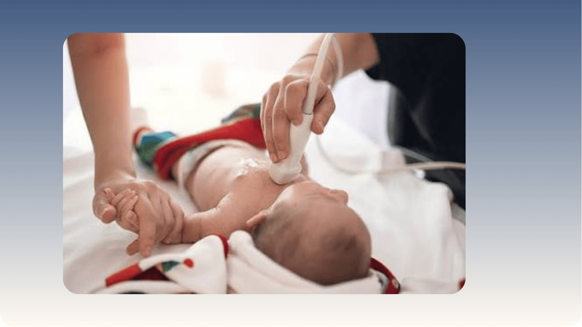
In my guide to POCUS in paediatrics, I described how bedside ultrasound has changed the way I diagnose and treat children across every age group, from infants with abdominal emergencies to children in respiratory distress. But neonatal POCUS deserves its own explanation, because the newborn, and particularly the premature newborn, is a patient population where the clinical challenges are unique, the stakes of delayed diagnosis are the highest, and the advantages of bedside imaging are the most compelling. A sick newborn may present with nothing more than a change in feeding behaviour, a subtle colour change, or a slight temperature instability. The same baby may have a serious lung problem, a cardiac condition, or a brain complication developing silently. As a Neonatologist and the clinician who introduced POCUS to paediatric practice in Eastern Uttar Pradesh, neonatal POCUS is one of the most important tools I have for getting inside a sick newborn's clinical picture in real time, without radiation, without moving the baby, and without waiting.
Why Newborns Cannot Wait for Formal Imaging
The practical constraints of imaging a sick newborn in a conventional radiology setting are significant. A premature infant on respiratory support, with intravenous lines, monitoring leads, and a temperature-controlled environment, cannot simply be disconnected and carried to a radiology suite without risk. The stress of transport, even a short distance within a hospital, can cause oxygen desaturation, hypothermia, and haemodynamic instability in the most vulnerable patients. A portable chest X-ray can be brought to the cot, but it provides limited information about cardiac function, fluid status, or brain integrity, and every X-ray in a preterm infant contributes to a cumulative radiation dose that clinicians prefer to minimise. POCUS at the cot side provides immediate, real-time answers to specific clinical questions without removing the baby from its monitored, temperature-controlled environment and without any radiation exposure. For families sitting beside their newborn's incubator in the neonatal unit, watching a doctor scan their baby at the cot and explain what the scan is showing in real time is also a form of communication that waiting for a radiology report simply cannot match.
Lung POCUS in the Newborn: Diagnosing Breathing Problems at the Bedside
Respiratory distress is the most common reason a newborn requires admission to a neonatal unit. The causes range from transient tachypnoea of the newborn, which resolves on its own, to respiratory distress syndrome from surfactant deficiency in premature infants, to pneumonia, pneumothorax, or pleural effusion. Distinguishing between these causes changes the management completely: a preterm infant with respiratory distress syndrome needs surfactant therapy, while a pneumothorax needs immediate decompression, and the two conditions can look similar clinically in the first minutes of life. Lung POCUS in the newborn identifies the specific ultrasound pattern associated with each of these conditions. The absence of normal lung sliding on one side, combined with the presence of a lung point, is a reliable sign of pneumothorax. A pattern of dense B-lines replacing the normal lung appearance indicates respiratory distress syndrome or fluid accumulation. Consolidation with air bronchograms indicates pneumonia. In a premature infant deteriorating in the first hours of life, a lung POCUS performed at the cot takes approximately two minutes and can direct the entire management decision.
[H2]
Cardiac and Cranial POCUS: The Two Most Critical Neonatal Applications
Cardiac POCUS in the newborn provides immediate information about cardiac function, heart structure, and the presence of pericardial effusion. A newborn with suspected congenital heart disease, persistent pulmonary hypertension, or shock from any cause can be assessed at the bedside within minutes. Whether the heart is contracting normally, whether there is fluid compressing the pericardium, and whether the right-sided pressures suggest pulmonary hypertension are all assessable with a focused neonatal cardiac scan, providing information that guides the immediate management while more comprehensive echocardiography is arranged.
Cranial POCUS in newborns takes advantage of the anterior fontanelle, the soft spot on the top of the skull that remains open in the first weeks of life. This acoustic window allows direct ultrasound assessment of the brain without X-ray or CT radiation. In premature infants, where the blood vessels of the germinal matrix are fragile and vulnerable to rupture under haemodynamic stress, intraventricular haemorrhage (IVH) is a serious complication that produces blood in the fluid-filled spaces of the brain. Left undetected, it can cause brain damage, hydrocephalus, and long-term developmental consequences. Serial cranial ultrasound through the fontanelle, performed at the cot side on the preterm infant's designated screening schedule, detects IVH at its earliest stages, grades its severity, and monitors for evolution over the days and weeks of the neonatal admission. At Vatsalya Children's Hospital, Varanasi, cranial POCUS for premature infants is part of the routine neonatal care protocol, allowing every premature baby at risk to be monitored without transport and without radiation.
To book a neonatal or paediatric consultation with Dr. Chhaya Pathak at Vatsalya Children's Hospital, Mahavir Road, Orderly Bazar, Varanasi, Uttar Pradesh 221002, call +91 9838585111 or visit linqmd.com/doctor/chhaya-pathak
Written by Dr. Chhaya Pathak, MBBS, MD (Paediatrics), Senior Consultant Paediatrician, Neonatologist and Intensivist, Pioneer of Paediatric POCUS in Eastern Uttar Pradesh, Co-Founder, Vatsalya Children's Hospital, Mahavir Road, Orderly Bazar, Varanasi, Uttar Pradesh 221002. 40+ years. 1,00,000+ patients. Phone: +91 9838585111.
Related reading
POCUS in Paediatrics: How Bedside Ultrasound Is Changing the Way Children Are Diagnosed and Treated
Pediatric Critical Care: Early Signs, Emergency Management, and Life-Saving Treatments for Children

Add a Comment